
Central serous retinopathy can make the centre of a page look blurred or bent, even when the edges seem clearer. A face may appear slightly different in size, or colours may look less bright in one eye. These changes can be worrying, especially when they fluctuate from one day to the next. This guide explains what central serous retinopathy is, what an eye specialist may check and why follow-up still matters when sight begins to improve.
What the name describes
Central serous retinopathy, also called central serous chorioretinopathy, affects the retina at the back of the eye. The macula is the small central area that helps you see fine detail. In this condition, fluid collects underneath the macula and makes it swell. That can change the central picture your eye sends to the brain. The condition usually affects one eye, but both eyes can be involved.
The name is not something you can confirm from symptoms alone. Several eye conditions can cause blur, distortion or a blank patch. An eye examination is needed to understand what is happening and where the fluid is. If you notice a change, describe it to an eye-care professional rather than trying to match it to a picture online. The RNIB guide to central serous retinopathy gives further background on symptoms and follow-up.
Changes you may notice
People can experience different combinations of symptoms. Central vision may look blurred, wavy or uneven. A straight edge, such as a door frame or line of text, may seem bent. A small blind spot can make a letter or part of a face difficult to see. Colours may seem duller, or an object may appear smaller than it really is.
Some people find that vision changes during the day. A symptom that seems better in the morning may feel more obvious later, or the reverse. This variation does not tell you whether the retina is healing. Keep follow-up appointments even if a good day makes the problem feel less urgent.
To explain the change clearly, cover one eye at a time only if this is comfortable, and compare the same page or object from the same distance. Do not press on the eye. Note what looks different, when you first noticed it and whether it is changing. That record can help the clinician understand your experience, but it is not a test for diagnosing yourself.
Why it can happen
Often, doctors cannot identify a single cause. RNIB describes the cause as unknown in many cases, while noting factors that may be linked with a higher chance of the condition for some people. These include steroid medicines, pregnancy, sleep apnoea, untreated high blood pressure, Cushing syndrome and H. pylori infection. The presence of any one factor does not prove that it caused a person’s eye problem.
Stress hormones have been discussed as one possible part of the picture. They do not explain every case, and the condition is not a personal failure or a sign that someone simply needs to relax. Avoid blaming yourself or another person for it.
If you use steroid medicine, do not stop or change it on your own. Steroids can be prescribed for important reasons and stopping suddenly may be unsafe. Tell both the clinician who prescribed it and the eye specialist about the medicine, including tablets, inhalers, creams, injections or eye drops where relevant. They can decide whether any change is appropriate.
What an examination can show
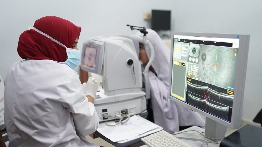
An eye specialist will ask when the change began and what it looks like. They may examine the retina and take images to check the macula. The exact tests depend on the clinical situation. An appointment is also a chance to discuss your other eye conditions, medicines and general health, without assuming that any one of them is the cause.
People living with diabetes may already have a routine for retinal checks. That is useful, but it does not replace an assessment of a new symptom. Our guide to diabetes and yearly retina checks explains why follow-up matters even when you feel well.
Before the appointment, write down your questions. You might ask which part of the retina is affected, whether the other eye needs checking, what changes should prompt an earlier call and when the next review should be. If a medicine may be relevant, bring its name or a list from your pharmacy. These details make the discussion more useful.
Why observation may be the first step
Many people improve without a procedure. RNIB says sight often improves over several months, commonly within three to six months, and that many people do not need treatment. A clinician may monitor the retina while it recovers. This is active follow-up, not neglect. Repeated checks help the team see whether fluid is settling, persisting or returning.
Recovery varies. A calendar estimate cannot predict what will happen in one person’s eye. Some cases last longer or return, and prolonged fluid can put sight at greater risk. Contact the clinic if your symptoms change in a way that concerns you or if you are unsure whether to wait for the next appointment.
Keep a simple record between visits. Note the date, which eye seems affected and whether lines look straight or distorted. Do not spend hours testing your vision or repeatedly checking small details. A brief note is enough to help you describe a change without turning the day into a series of self-tests.
When treatment may be discussed
If fluid does not improve or the condition keeps returning, an ophthalmologist may discuss treatment. RNIB describes thermal laser and photodynamic therapy as options that may be considered in some cases. The choice depends on details such as how long the fluid has been present, whether it has recurred and where the leak lies in relation to the centre of the macula.
Not every treatment is suitable for every leak. RNIB notes that thermal laser cannot be used when the leak is too close to the centre because treatment could cause more harm than benefit. The aim of treatment is to reduce or resolve fluid. It cannot restore retinal cells that have already been damaged.
Ask the specialist to explain the expected benefit, limits, risks and alternatives for your own scan. If a procedure is suggested, it is reasonable to ask what would happen if the team continued monitoring instead. A clear answer should relate to the findings in your eye, not a promise that vision will return to a particular level.
Making daily tasks easier
While central vision is unsettled, small adjustments can reduce frustration. Increase the text size on a phone or computer. Use even room lighting and choose a surface with good contrast. If a label is difficult to read, use a larger version or ask someone you trust to check it. These changes do not treat the retina, but they may make a task easier while you wait for clinical advice.
If one eye has much poorer sight, take care when pouring hot drinks, using stairs or moving through a crowded place. Keep frequently used objects in consistent locations. If reading remains difficult despite glasses, ask the eye team whether a low-vision assessment or magnifying aid may help.
For a broader explanation of the photographs and dilating drops used in some eye checks, see what a dilated eye examination checks. That guide is general information; the clinician will choose tests for your own symptoms.
When to seek advice
Arrange an eye assessment when you notice a new distortion, blur, blank patch or change in colour vision. Do not wait for symptoms to become severe before asking for advice. If your sight changes suddenly or worsens quickly, contact an eye-care service promptly and explain that the change is new.
This page is general education, not a diagnosis. It cannot tell whether your symptoms are central serous retinopathy or another condition. An eye specialist can check the retina, discuss your individual situation and decide how soon you should be reviewed.
Questions to take to your review
- What did the examination or scan show about the macula?
- What changes should make me contact the clinic before my next appointment?
- Could any of my current medicines be relevant, and who should I speak with before changing them?
- When should the next review happen if my sight improves or stays the same?
- Would lighting, magnification or another practical aid help with a task I am finding difficult?
Bring a family member if that helps you remember the answers. Ask the clinician to write down the next appointment date and the contact route for new symptoms. If an instruction is unclear, ask for it to be explained in everyday words before you leave.
Where to turn
If you would like to discuss arranging an eye examination, use the PEBS Hospital contact page. It does not confirm which tests or treatments may be available for an individual case. If you wish to support eye care, information is on the donate page. For medical advice about new or changing vision, speak with a qualified eye-care professional.
Have a question about your eyes?
PEBS Hospital in North Karachi can examine you and explain your options. Call the helpline on 021-36908053, or send a message and the team will reply.
This article is general information and does not replace an examination by an eye doctor.