
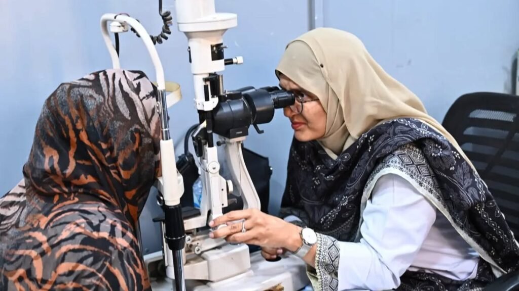
Retinal vessel occlusion means a blood vessel has become blocked at the back of the eye. It can cause a painless change in sight. If all or part of your vision suddenly disappears, seek emergency care immediately. Do not wait for pain, and do not wait for a routine appointment. This guide explains the difference between artery and vein blockages using RNIB’s patient information.
Why the type of vessel matters
Arteries bring oxygen-rich blood to the retina. Veins carry used blood away. The retina needs a continuing supply of blood to function. A blockage on either side can damage sight, but the process and treatment are different.
An artery blockage interrupts the supply reaching the tissue. A vein blockage prevents blood from draining normally. Blood and fluid can then leak into the retina, causing bleeding and swelling.
Both types usually affect one eye at a time and can be painless. The amount of sight affected depends partly on where the blockage occurs. A small branch and a central vessel do not supply or drain the same area.
Sudden loss of vision is an emergency
A retinal artery occlusion can cause loss of sight within seconds. RNIB advises immediate attendance at an emergency department for sudden loss of all or part of vision. This applies even when only one eye seems affected.
Vision can also disappear briefly and then return. This can be called amaurosis fugax. It may indicate a temporary interruption to blood flow. The fact that sight returned does not make the event safe to ignore.
Not every temporary visual loss has the same cause. Immediate investigation is needed to find out what happened. Tell the medical team when it began, how long it lasted and which eye or part of vision was affected.
Artery blockages and their names
The central retinal artery divides into branches after entering the eye. A blockage in the central artery can affect sight across the eye. A blocked branch generally affects the area served by that branch.
CRAO means central retinal artery occlusion. BRAO means branch retinal artery occlusion. These abbreviations may appear in a hospital letter. Ask the doctor to explain which vessel is involved and what that means for your findings.
Retinal cells are very sensitive to loss of oxygen. Damage can become permanent. Emergency assessment matters even though there is no guaranteed treatment that restores sight after an artery occlusion. Do not try to manage the problem at home.
Why arteries can become blocked
Fatty deposits can build up inside artery walls. This process, called atherosclerosis, narrows the space for blood to flow. A piece of material can also travel from a larger vessel and lodge in a smaller one.
That travelling material is called an embolus. If it blocks a retinal artery, oxygen cannot reach part of the retina normally. Less common causes include inflammation of vessel walls, injury and certain blood disorders.
Looking for the cause is part of care after the eye emergency. The problem may have implications beyond the affected eye. A doctor may arrange investigations and involve other specialists according to the findings.
How a vein blockage may feel
A retinal vein occlusion may cause dimming or blurring over hours or days. The change may involve part or most of the affected eye’s sight. Any new visual change needs an examination.
If sight deteriorates markedly or quickly, go to an emergency department promptly. Do not attempt to distinguish an artery blockage from a vein blockage using the speed alone. That distinction needs an assessment.
CRVO refers to a central retinal vein occlusion. BRVO refers to a branch retinal vein occlusion. A central blockage usually affects more of the retina. A branch blockage may affect a smaller area and sometimes improves as swelling settles.
Treatment after a vein occlusion
RNIB explains that treatment addresses complications of a vein occlusion rather than removing the blockage itself. Some people need monitoring initially. Others need treatment for swelling, abnormal blood vessels or raised eye pressure.
A decision to monitor does not mean the event was unimportant. The eye team is watching for changes that could require treatment. Follow-up can continue for a substantial period even when no procedure is needed at the first appointment.
Ask what the doctor is monitoring, when you should return and what symptoms should prompt earlier contact. Keep the plan somewhere you and a relative can find it. A clear plan is easier to follow than a general instruction to come back later.
Swelling at the centre of the retina
Fluid can collect in the macula, the central area used for detailed vision. This is called macular oedema. It can make reading, seeing faces and watching television difficult.
Depending on the findings, treatment may include anti-VEGF or steroid injections. Mild swelling may sometimes be observed because it can settle. The eye specialist decides whether treatment is needed and when it should begin.
Ask which complication a proposed injection is treating. Also ask how response will be checked. The name of the original blockage does not, by itself, tell you the number of visits or treatments you will need.
New vessels and eye pressure
When areas of the retina lack oxygen, the eye may grow abnormal new blood vessels. This is called neovascularisation. These vessels are fragile and can bleed. Most people with a vein occlusion do not develop this complication.
New vessels can also grow near the eye’s drainage area. They may interfere with fluid drainage and raise eye pressure. If pressure damages the optic nerve, glaucoma can result.
Laser treatment may be used for abnormal vessel growth. Drops or other treatment may be needed for raised pressure. This is another reason to attend reviews even when the initial blur seems unchanged.
Checking general health is part of eye care
Risk factors include high blood pressure, high cholesterol, diabetes, smoking and certain clotting disorders. Raised eye pressure is also relevant. Some people have no identifiable risk factor after investigation.
The doctor may recommend checks of blood pressure, blood sugar and cholesterol. After an artery occlusion, further investigation may include the arteries in the neck. The exact tests depend on the medical assessment.
These checks cannot reverse retinal damage that has already occurred. They can help identify risks needing treatment. For people with diabetes, our guide to diabetes and retinal checks explains why eye care belongs alongside general medical care.
Habits and medicines after the diagnosis
Stopping smoking, eating a balanced diet and maintaining physical activity can support blood vessel health. Ask your clinician about suitable activity and help with smoking cessation. Advice should account for your wider health and abilities.
Take prescribed medicines according to your own plan. If there is a problem with a medicine, ask the prescribing doctor or pharmacist. Do not start a blood thinner or change treatment because someone else received it after an eye problem.
Ask which clinician will review each risk factor. Eye follow-up and general medical follow-up have different purposes. Knowing who is responsible makes it easier to keep both parts of care moving.
Adjusting to a change in one eye
Using the eyes together helps with judging distance. After a change in one eye, steps, kerbs or reaching for objects may feel less reliable. Adjustment can take time.
Tell the team about practical difficulties, including reading or moving around. If both eyes have reduced vision, a low vision assessment may explore magnification, lighting and contrast. Support is still worthwhile when sight cannot be fully restored.
Ask for individual advice before returning to driving. The ability to manage familiar tasks at home does not establish safe driving vision. Overseas licensing rules described in a source do not replace local requirements.
Where to take questions
The source for this guide is RNIB’s retinal vessel occlusion information. To make a non-emergency enquiry about your case, use the PEBS enquiry page for retinal vessel occlusion. Those wishing to give can use the donate page.
For sudden sight loss, the next step is emergency assessment now, even if the eye looks normal and does not hurt.
Have a question about your eyes?
PEBS Hospital in North Karachi can examine you and explain your options. Call the helpline on 021-36908053, or send a message and the team will reply.
This article is general information and does not replace an examination by an eye doctor.